Student projects (závěrečné práce a projekty)
Our department offers a wide range of student projects (B.Sc./Bc. and M.Sc./Ing.). Please see below some suggested project topics. Also note that this is an open list of projects, by which we mean that it is possible to customize the projects with respect to student’s goals and abilities. If you, however, feel that none of the projects suits you but you would like to still work on a project/write a thesis on machine learning/(medical) image processing/data mining/assistive technologies then write an email to the corresponding researcher and we will soon get back to you and we will discuss possible solutions.
Automated Brain Extraction
Type: Bc. or Ing. individual or diploma project
Supervisor: Ing. Milan Němý, Ph.D. (milan.nemy@cvut.cz, nemymila@fel.cvut.cz)
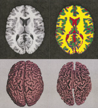
Accurate segmentation of brain and non-brain tissue is a crucial step of many applications in brain imaging. For example, all volumetric studies begin with brain extraction of structural magnetic resonance (MR) images of the head to estimate the subject’s intracranial volume which is later used for normalization. Manual brain/non-brain segmentation typically takes between 15 min and 2 hr per 3D volume and requires sufficient training. It is, therefore, no wonder that automatic methods are very popular. In this project, you will report on various techniques to tackle this problem and implement one of the methods. Then, you will evaluate its accuracy and compare it with existing tools.
Tasks:
- Study classical and modern approaches to the problem of (automated) brain extraction in MRI data.
- Implement a selected method of automated brain extraction in MATLAB.
- Compare the accuracy of the implemented method with publicly available automatic brain extraction tools using a golden standard model.
- Assess the strengths and weaknesses of your implementation of the above-chosen method to other extraction techniques.
Diffusion Tensor Tractography in Alzheimer’s Disease
Type: Bc. or Ing. individual or diploma project
Supervisor: Ing. Milan Němý, Ph.D. (milan.nemy@cvut.cz, nemymila@fel.cvut.cz)
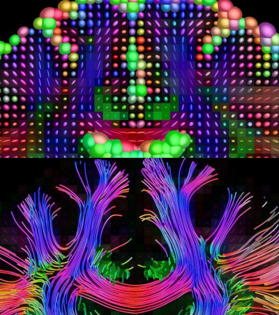
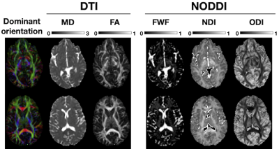
Alzheimer’s disease (AD) is the most common form of senile dementia which is generally characterized by memory loss followed by a progressive decline in other cognitive domains. Several recent studies have proposed that cognitive decline in AD is a consequence of disruptions in the structural and functional connections between brain regions. Diffusion-weighted MRI (DWI), a variant of standard anatomical magnetic resonance imaging (MRI), is sensitive to microscopic white matter injury not always detectable with standard anatomical MRI. DWI tracks anisotropic water diffusion along axons, revealing microstructural white matter bundles in the brain’s anatomical network. In this thesis, you will learn about various techniques for tracking white matter bundles using DWI data and you will implement one of the algorithms in MATLAB. Next, you will be asked to use your implementation to find several commonly identified tracts, evaluate their integrity and subsequently compare it between healthy controls and the AD group.
Tasks:
- Study selected literature on diffusion tensor imaging, several tractography algorithms, and changes in brain connectivity in Alzheimer’s disease (AD).
- Implement a selected tractography algorithm in MATLAB.
- Investigate differences in brain connectivity between healthy controls and the AD group using your implementation of a tractography algorithm.
- Assess the strengths and weaknesses of your implementation of the above-chosen method to other tractography techniques.
Diffusion Tensor Imaging in Alzheimer’s Disease
Type: Bc. or Ing. individual or diploma project
Supervisor: Ing. Milan Němý, Ph.D. (milan.nemy@cvut.cz, nemymila@fel.cvut.cz)
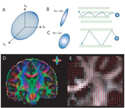
Alzheimer’s disease (AD) is the most common form of senile dementia, generally characterized by memory loss followed by a progressive decline in other cognitive domains. Several recent studies have proposed that cognitive decline in AD is a consequence of disruptions in the structural and functional connections between brain regions. Diffusion-weighted MRI (DWI), a variant of standard anatomical magnetic resonance imaging (MRI), is sensitive to microscopic white matter injury not always detectable with standard anatomical MRI. DWI tracks anisotropic water diffusion along axons, revealing microstructural white matter bundles in the brain’s anatomical network. In this project, you will learn about a technique for reconstructing a model of white matter diffusivity, and you will implement one of the algorithms in MATLAB. Next, you will be asked to use your implementation to compare white matter integrity between healthy controls and the AD group.
Tasks:
- Study selected literature on diffusion tensor imaging (DTI) and changes in brain connectivity in Alzheimer’s disease (AD).
- Implement DTI model reconstruction in MATLAB.
- Investigate differences in brain connectivity between healthy controls and the AD group using your implementation of DTI.
- Assess the strengths and weaknesses of your implementation.
Classical Automated Lesion Segmentation in Magnetic Resonance Imaging in Multiple Sclerosis
Type: Bc. or Ing. individual or diploma project
Supervisor: Ing. Milan Němý, Ph.D. (milan.nemy@cvut.cz, nemymila@fel.cvut.cz)
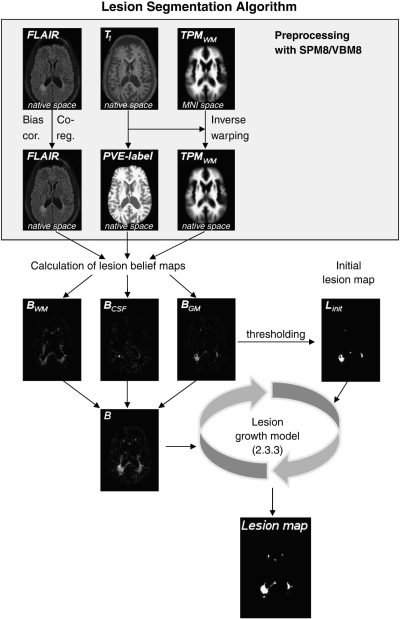
Magnetic resonance imaging (MRI) represents one of the most important tools for diagnosing multiple sclerosis, monitoring the disease progression, and predicting its future development. One of the leading indicators of disease development is the number, location, and volume of brain lesions caused by the demyelinating process. Traditionally, T2-weighted sequences and FLAIR imaging are used to detect these hyperintense lesions. However, a variety of new MR imaging techniques are currently beginning to be used to increase detection sensitivity and provide a more comprehensive view of central nervous system damage. In this project, the student will create an overview of MR imaging techniques for the segmentation of brain lesions in multiple sclerosis. Furthermore, the student will implement selected classical segmentation procedures and will compare their accuracy with available segmentation tools.
Tasks:
- Study of traditional and modern MRI techniques for imaging and segmentation of MS brain lesions.
- Implement three selected automatic brain lesion segmentation algorithms (in Matlab or a similar environment).
- Compare the accuracy of your implementation with standard brain lesion segmentation tools.
- Assess the strengths and weaknesses of your implementation of the above-chosen method to other brain lesion segmentation techniques.
Deep Learning-Driven Automated Lesion Segmentation in Magnetic Resonance Imaging in Multiple Sclerosis
Type: Bc. or Ing. individual or diploma project
Supervisor: Ing. Milan Němý, Ph.D. (milan.nemy@cvut.cz, nemymila@fel.cvut.cz)
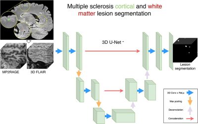
Magnetic resonance imaging (MRI) represents one of the most important tools for diagnosing multiple sclerosis, monitoring the disease progression, and predicting its future development. One of the leading indicators of disease development is the number, location, and volume of brain lesions caused by the demyelinating process. Traditionally, T2-weighted sequences and FLAIR imaging are used to detect these hyperintense lesions. However, a variety of new MR imaging techniques are currently beginning to be used to increase detection sensitivity and provide a more comprehensive view of central nervous system damage. In this project, the student will create an overview of MR imaging techniques for the segmentation of brain lesions in multiple sclerosis. Furthermore, the student will implement a selected segmentation procedure using a deep learning approach and will compare their results with a gold standard.
Tasks:
- Study of traditional and modern MRI techniques for imaging and segmentation of MS brain lesions.
- Implement an automatic brain lesion segmentation routine using a deep learning approach (in Matlab or a similar environment).
- Compare the outcomes of your implementation to manually segmented data.
- Assess the strengths and weaknesses of your implementation of the above-chosen method to other brain lesion segmentation techniques.
Voxel-based Cortical Thickness Measurements in MRI
Type: Bc. or Ing. individual or diploma project
Supervisor: Ing. Milan Němý, Ph.D. (milan.nemy@cvut.cz, nemymila@fel.cvut.cz)
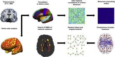
The proposed Bc./Ing. thesis will focus on the analysis of cortical thickness and its implications on brain connectivity and network resilience. Cortical thickness, the measurement of the brain’s outer layer, is a crucial indicator of neural health and cognitive function. This thesis will guide students through the process of developing their own computational tools for cortical thickness analysis, despite the availability of existing tools, to ensure a deep understanding of the underlying methodologies.
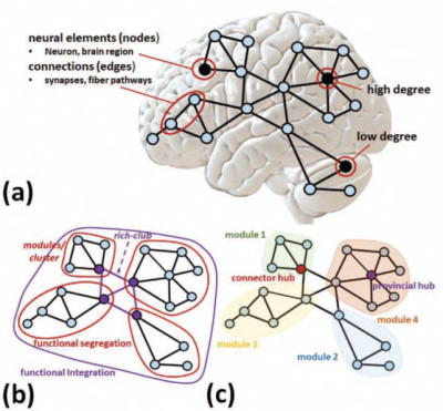
Students will begin with the preprocessing of T1-weighted MRI data (magnetic resonance imaging), followed by parcellation to define cortical regions. They will compute inter-regional correlations of cortical thickness to create a binarized connectivity matrix. The next phase involves employing graph theory to analyze these matrices, where students will simulate random failures and targeted attacks to assess network resilience. This analysis aims to uncover hidden patterns and relationships between different brain regions, providing insights into how cortical thickness variations influence brain connectivity.
Through this thesis, students are expected to gain proficiency in MRI data processing, parcellation techniques, and graph theory applications. They will be required to implement custom solutions for each step, culminating in a comprehensive comparison of graph measures to determine the impact of cortical thickness variations on neural network integrity.
Analysis of Brain Connectivity Changes in Alzheimer's Disease Using Diffusion Magnetic Resonance Imaging
Type: Bc. or Ing. individual or diploma project
Supervisor: Ing. Milan Němý, Ph.D. (milan.nemy@cvut.cz)
The quality of structural brain connectivity can be non-invasively measured using diffusion magnetic resonance imaging (dMRI) methods. In particular, tractography, which allows for a detailed examination of the connections between different regions of the cerebral cortex and subcortical structures, represents a key tool for analyzing how efficiently and effectively different parts of the brain are interconnected. These methods enable the study of not only local connections but also the organization of entire brain networks, which play a crucial role in executing specific functions. Furthermore, the analysis of such networks provides valuable insights into how they change in neurodegenerative diseases.
As part of this work, the student will create an overview of techniques for analyzing local brain connections and brain networks using dMRI. Additionally, the student will independently implement a selected tractography method on a provided set of patient imaging data and use it to compare the quality of brain connections in patients with Alzheimer’s disease versus healthy controls. The results will then be compared with the available literature.
Development of an Imaging Browser for Diffusion MRI Analysis
Type: Bc. or Ing. individual or diploma project
Supervisor: Ing. Milan Němý, Ph.D. (milan.nemy@cvut.cz)
Modern medical imaging research relies on efficient tools for visualization, processing, and analysis of MRI data. Advanced diffusion MRI (dMRI) techniques such as diffusion tensor imaging (DTI), diffusion kurtosis imaging (DKI), restriction spectrum imaging (RSI), and others provide deeper insights into microstructural changes in brain tissues. However, existing imaging tools are often either too complex or lack support for specialized dMRI models.
In this project, you will develop a user-friendly front-end imaging browser in Python for visualizing and analyzing diffusion MRI data. The application will enable loading, displaying, and processing dMRI-derived maps and segmentations. The project will focus on usability, integration with existing imaging pipelines, and facilitating efficient visualization of diffusion metrics.
Modelling Diffusion Signal for Glioma Characterization Using Advanced DWI Methods
Type: Bc. or Ing. individual or diploma project
Supervisor: Ing. Milan Němý, Ph.D. (milan.nemy@cvut.cz)
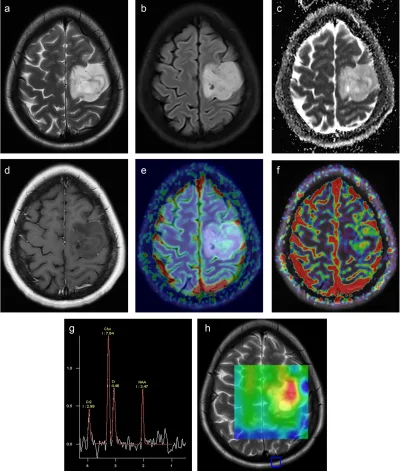
Brain tumors, like gliomas, can be difficult to classify using standard MRI scans. Doctors often use a technique called Diffusion-Weighted Imaging (DWI) to measure how water moves in brain tissue. This helps them understand if a tumor is aggressive (high-grade) or slow-growing (low-grade). A simple DWI measure called Apparent Diffusion Coefficient (ADC) is commonly used, but it is not always accurate. Newer advanced diffusion models—such as Diffusion Kurtosis Imaging (DKI), Restriction Spectrum Imaging (RSI), and Vascular Models (VERDICT)—can give better results by analyzing how water moves in different ways inside tumors.
In this project, you will learn how these advanced MRI methods work and use them to improve tumor classification. You will test different mathematical models on real patient data and see how well they can predict whether a tumor is high-grade or low-grade. You will also try machine learning techniques to automatically classify tumors and check if they have a specific genetic mutation (IDH mutation), which affects treatment options.
Using of HL7 Infobutton standard
Type: Bc. or Ing. individual or diploma project
Supervisor: Ing. Michal Huptych, Ph.D. (Michal.Huptych@cvut.cz)
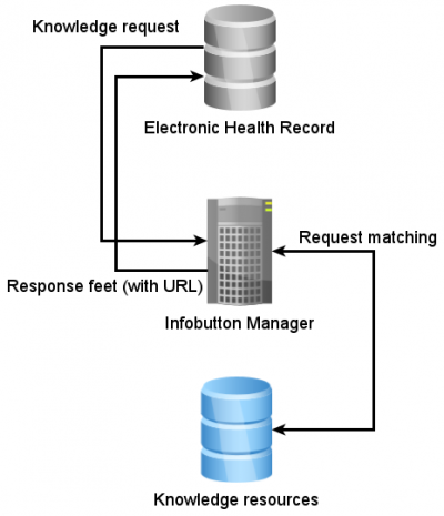
The HL7 is mainly known as a communication standard for exchanging medical data. However, the HL7 is a standard organization that cover a far more extensive area of technologies than only exchange data. From the perspective of expert systems, the one of interest is HL7 Infobutton (https://wiki.hl7.org/Product_Infobutton). Briefly, the Infobutton is a tool for the distribution of knowledge and evidence from the different knowledge sources to the clinical information system. The approach should ensure a simple way how a physician can reach evidence like articles, guidelines or results of models. There is one implementation of the HL7 Infobutton, called Open InfoButton, that is developed in Java. Therefore, the preferred programing language of this project is also Java. The aim of this project is to deploy the Open Infobutton system and develop the environment for knowledge and evidence distribution.
Tasks:
- Study problematics of knowledge and evidence distribution in clinical practice, especially standard HL7 Infobutton
- Study project Open Infobutton (https://www.openinfobutton.org/) and deploy the project.
- Define possible knowledge and evidence resources and a way how to implement those including into HL7 Infobutton/ Open Infobutton Project
- Implement technology HL7 Infobutton into a small information system (called Electronic delivery Book) used at the University Hospital Brno.

